Monday 8 July 2013

Classification

Classification is not a scientific theory.  It is the effort of one species (us) to try and group organisms in such a way as to make sense to us.

The way in which we will classify is by using dichotomous keys.  These leave us with 2 choices. If structured well, it can be a simple yes/no choice.  A flow chart arrangement can be seen below.

Dichotomous Key Structure

Another option is where the choices are arranged in a table.  This example is shown below.


The advantages of keys are:

  1. Allows for consistency when classifying
  2. Can be used by a non expert

The disadvantages of keys are:
  1. If not well written can cause confusion
  2. If an organism is new to science, it may be difficult to assign or mistakenly classified


The father of classification is Linneaus. He named organisms using a binomial system, giving the genus name first, then the species name.  In the name, there should be some indication of the characterisics.  For example the name Panthera leo (for the lion) indicated "big cat, lion).


As time went on, and the number of plants and animals described increased, it became obvious that the system of 2 names was not going to be sufficient. The invention of the microscope also meant that the microscopic world was now open.  Eventually the groups of organisms were organised into 5 kingdoms according to their key features at the cell level.


Picture


However, the advance of technology and the uncovering of the Archaea has led to major revision of the classification system. Rather than having 5 kingdoms, its now proposed that 3 domains be placed at the top classification level.  4 of the 5 kingdoms are swept into one domain and the remaining one is divided into 2.


Archaea and Eubacteria


With the advent of better technology (particularly DNA sampling and sequencing), bacteria were being grouped into 2 large groupings.  These were the Archaea and the modern bacteria (referred to as Eubacteria).  Members of the Eubacteria were better known because they had grown on suitable mediums for nearly a century.  The link between bacteria and disease was one of the reasons this field was established.


The major features of Eubacteria are as follows:

1. Generally able to survive in the presence of oxygen
2. Generally they have two types of cell walls, that can be identified by certain staining techniques.
3. Widely distributed in a vast number of environments 
4. Essential in nutrient cycles as decomposers.


The major features of Archaea are as follows:

1. Often found in extreme environments
2. Many cannot grow in the presence of high oxygen levels
3. Cells walls are chemically very different from Eubacteria
4. DNA sequences very different from Eubacteria
5. A high proportion of these are autotrophic.  


Some of these bacteria live in environments thought to be like early Earth (hot, volcanic, chemical rich) it is possible that Archaea were the ancestors to many cell types found today. 

Bubbling mud at Tokaanu Hot Springs


Technology and Understanding of Prokaryotes


One of the technologies that can be credited with a recent surge in understanding of bacterial cell types can be credited with the deep sea submersible ALVIN.  When it explored the hydrothermal vents along the mid-Atlantic ridge, it not only sampled larger organisms that were found but also bacterial mats that were on the surfaces on the surrounding rocks.


It was found that these bacteria were vastly different from those that were currently identified.  This led to scientists sampling bacteria in other extreme environments such as hot springs, acid pools and saline lakes.  Again, these bacteria are very different.

Other technologies that were utilised were DNA sequencing and the use of a technology called the Polymerase Chain Reaction.  Using this scientists have found varieties of bacteria that they haven't been able to grow in culture.  Amazingly a teaspoon of soil contains up to 10000 individual species of bacteria!

When the DNA sequences of the bacteria from extreme and often anaerobic conditions were compared to others that more was known about, it was found that they were vastly different.  

Given the incredible numbers and diversity of bacteria and the differences between them, this has led to a revision of the 5 kingdom system of classification.  More on this later.

Another piece of technology that should not be overlooked is the electron microscope.  It showed the internal workings of the cells, the structure of bacterial cell walls, and in some cases showed that bacterial cells formed tough resilient spores that could withstand tough environments.


Sunday 7 July 2013

The first cells, chemoautotrophs and photoautotrophs

The Urey Miller experiment caused a great deal of excitement and was partially responsible for launching a whole new branch of biology called exobiology.

Variations of the Urey-Miller experiment were soon trialed.  It was found that sugars could be produced, lipids that form bilayers could be formed, proteins could be assembled, and molecules that self replicate could be made.  However, how these were able to be aggregated into something resembling a cell is still a matter of conjecture.  However, it is agreed that first cells appeared on Earth between 3.5 to 3.8 billion years ago.  The first fossil evidence is dated to 3.45 billion years ago.


Bacterial specimens found in rocks from the Barbeton Mountains, South Africa.  The rocks are dated to over 3.4 billion years old.
  

It is thought that the first cells were likely to be heterotrophic. That is, they consumed the complex energy yielding chemicals that were in the environment.  This would have been limited and would have run out fairly quickly.  Varieties of bacteria that were able to have produced their own food (autotrophs) would have survived while most of the heterotrophs would have perished.  These bacteria are likely to be the ancestors of what we now call the Archaebacteria.  These are found in extreme environments such as submarine vents in the deep oceans, hot springs, salt lakes etc.


These bacteria utilise fairly simple chemical compounds to obtain their own energy and complex substances needed for growth and division.  Most of these Archaebacteria are anaerobic, supporting the idea that they had their origins in an oxygen free environment.

From this group of bacteria, a new type arose. Rather than being powered from the chemicals produced by volcanoes, they utilised the sun to power the processes need to grow and divide.  These were the cyanobacteria, and their activities would change the Earth and the path life would take.


The first fossil evidence of photosynthesis were the appearance of fossilised stromatolites in WA dated to 3.4 billion years old. The largest colony of stromatolites in the world is currently in Shark Bay, also in Western Australia.
These are colonies of photosynthetic bacteria which form columns as they grow.  A by-product of their metabolism is oxygen.  These organisms were largely responsible for the removal of iron from the oceans, and a for adding large portion of the atmospheric oxygen and ozone layer.  For over 2 billion years, these bacteria were the dominant fossil found in the fossil record.

Thursday 6 June 2013

From single cells to multicellular organisms


In the process leading up to multicellular organisms, eukaryotic cells had to form.  A suggested mechanism is below.  It is thought that a progenitor cell may have got larger than other cells.  To maintain SA:vol ratios it folded its membranes, some of which then enclosed the DNA (forming a nucleus) and the endoplasmic reticulum and Golgi apparatus.


From here we move into something called endosymbiosis (endocytosis - to engulf something, often for nutrients, symbiosis - a long term interaction between 2 things, often to the point where one can survive without the other).

It is thought that the progenitor cell engulfed a heterotrophic bacteria and over time it degenerated into a mitochondria.  These cells were to become heterotrophic eukaryotes.  One of these also engulfed a photosynthetic prokaryote.  This photosynthetic prokaryote degenerated into a chloroplast and the entire cell was the ancestral plant cell.

Evidence to support this idea includes:

  1. The fact that mitochondria and chloroplast divide independently of each other
  2. The DNA of mitochondria and chloroplasts is circular (like bacteria) and has many common bacterial sequences


A link to describe the process in more detail is here.


Now that we have cells that are structurally similar to modern ones the next step is to get them to work cooperatively.  The levels of cooperation were thought to be 

One example is colonial algae,  Volvox.  It is broken into 2 cell types; cells for swimming (outer shell) and ones for reproduction (small green spheres)


In the case of the Portuguese Man O War (bluebottles)there are a number of additional cell types which are working cooperatively to assist the survival or the organism.  Just to clarify the labelling here, the Gonozooids (reproductive structures) are the light blue ones near the polyp while the Gastrozooids are the red/brown ones just below.


From here it is possible to see further increases in cooperation between cells, and therefore increasing complexity of the organisms that possess them.

The Urey Miller Experiment

In 1953, Stanley Miller, a PhD student, proposed to his supervisor Harold Urey, an experiment to test if the chemosynthetic origins of life were possible under the conditions of what early Earth was like.

This would in part answer the chicken and the egg question about whether it was possible for the early Earth to produce the chemicals needed to sustain life, before life itself actually got going on Earth.


Energy source and some of the gases that Miller proposed to use.           Note NO OXYGEN.

Miller then set up his experiment as follows:


Over a number of days the apparatus showed orange brown materials sticking to the glass and in solution.  Chemical analysis showed the following:


The final group of materials include amino acids (used to make proteins). Other substances include metabolites that certain types of cells can get energy out of.  


The findings were significant for a number of reasons:

  1. The early Earth could not have had oxygen present (supported by other geological evidence)
  2. The early Earth had conditions that could have allowed for the generation of molecules that would sustain life
  3. Other variations of the experiment showed that molecules that could be used in DNA and RNA could be produced.
So this experiment supports the chemosynthetic theory, but because we weren't there to observe it, it is NOT proven.


Monday 3 June 2013

Chemosynthesis versus Panspermia

There are 2 theories that are proposed to try and explain the origings of life.  These are:


  1. Chemosynthetic origins - The chemicals of life were made on Earth.  Once made, these eventually combined to make living organisms.
  2. Panspermia - The chemicals of life and/or life was seeded from asteroids and meteorites from space.

Lets start with chemosynthesis.

Looking at the Earth 3.8 billion years ago, it was thought to be highly volcanic, anoxic and had liquid water.  Lightning storms were thought to be frequent around erupting volcanoes (as they are today).  The surface of the planet was being bombarded with radiation as there was no ozone layer formed. The atmosphere was thought to be made up of simple molecules.


It was proposed that the energy of any of the above sources could have been used to break and reform chemical bond, thus turning the simple molecules of the atmosphere into more complex ones.  There will be some description about the experiments involved in this later.

Panspermia describes the idea that complex molecules form in space.  Many dust clouds in space contain carbon, nitrogen and water (ice crystals) in them. In addition asteroids and comets contain these elements as well.  Energized by radiation from the stars the chemical bonds can be rearranged to form complex materials some of which are found on Earth.


The conditions thought to occur in deep space have been modeled in Earth labs and so this theory is plausible as well.  If we find life of a similar chemical make up on other Earth-like planets, it may be that Panspermia becomes the preferred model.  Given what we currently know, it appears that the chemosynthetic origins of life is still favoured. The experiment that got the ball rolling on this was the Urey-Miller experiment performed in 1953. This is the subject of the next post.

Life on Earth - Conditions of Early Earth

This module looks at the development of life on Earth and the conditions that may have spawned  life.  To do this we need to see what the initial conditions of life on Earth were like.

4.5 billion years ago to 3.8 billion years ago.


At this time the Earth has no liquid water, and no stable crust.  The planet was still subject to heavy asteroid bombardment.  Because of the heat on the surface, any gases present would have escaped into space.  


3.8 - 3.45 billion years ago


By this time the Earth had cooled to the point where stable crust had formed and the temperature had cooled to the point that liquid water covered most of the surface of the planet.  Volcanic activity was still violent and frequent, but the gases being released from them were beginning to create an atmosphere.

This atmosphere was thought to be rich in methane, ammonia, carbon dioxide, carbon monoxide and possibly cyanide.  No free oxygen was present at this stage.  Hydrogen may have been formed from the spitting of water by UV radiation but the oxygen would have reacted quickly.  The lack of oxygen makes this atmosphere anoxic.

A number of small volcanic island were thought to have been forming at this time.  At the edges there may have been hot mud springs.  These springs may have been the place where life arose.  But before life can start, the complex molecules for life need to be created.  This is a topic for another post.

Tuesday 16 April 2013

Cell Division

This section is the last of the topic "Patterns in Nature".  Cell division is very important for organisms.  Certain cells quite literally wear out, others are damaged by the environment and others can be damaged or killed by some type of trauma.

In addition, organism growth is a result of cell division, not cell growth.  Remember what happens to cells when they get larger?

And finally, not all cells will divide for your entire life.  For example, nerve cells tend to stop dividing completely by about the age of twelve in humans.  This is why injuries involving nerves, the spine or the brain are often lifelong.

The type of cell division we are looking at in some detail is mitosis.  There are 2 important stages in cell division:


  1. Division of the DNA
  2. Division of the cytoplasm.
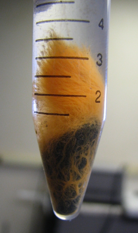
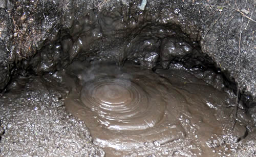
Starting with the DNA, when we see the nucleus, the DNA is essentially unwound so that the cell can use it for instructions for various cell processes and products.  It is a bit like the wool in the left hand picture here.  If we were to divide this up, chances are there would be large amounts of DNA breakage which will result in the cells being not viable.  To prevent this, the cell winds up the DNA (the process is called condensation) so that they form the visible chromosomes.  In doing so, they arrange the DNA into structures like similar to those on the right, which is much easier to divide into 2 cells.


Let's run through the stages of mitosis.

  1. Interphase - This is the longest phase and is the stage where DNA is copied and the cell increases in size.  Another feature of anaphase is mitochodria and chloroplasts divide independently of the cells at this stage.
  2. Prophase - The nucleus begins to condense into visible strands of DNA or chromosomes
  3. Metaphase - The chromosomes align themselves centre of the cell along the "equator"
  4. Anaphase - The chromosomes pull apart forming chromatids
  5. Telophase - The DNA in each daughter cell begins to unwind forming a new nucleus.


However, division of the nucleus is only one part of cell division.  The cytoplasm mus also be equally divided.  If its not, then the one of the daughter cells is likely to be non-viable, because there is not enough cytoplasm containing nutrients and organelles for the cell to keep functioning. If that happens, why divide in the first place?

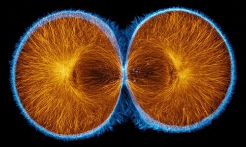
To ensure cytokinesis (division of the cytoplasm) takes place effectively, the cell forms a band of contractile proteins around the equator of the cell.  These pinch the cell into two fairly equal parts.


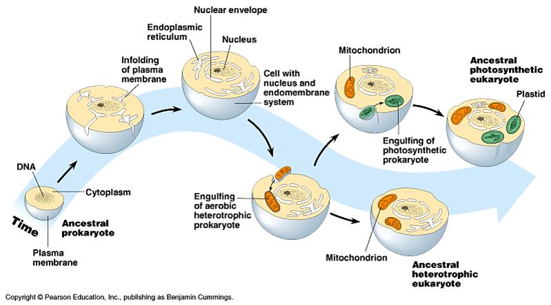
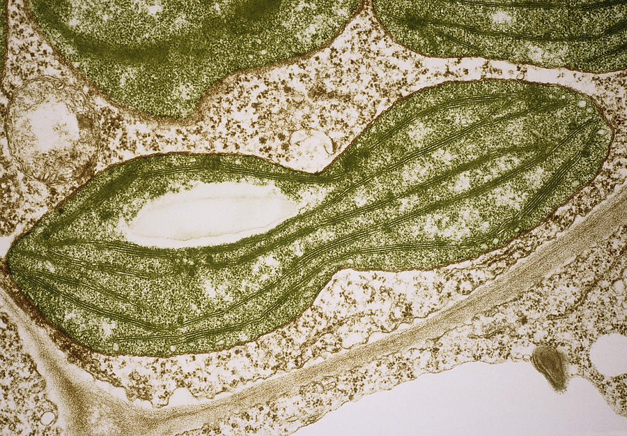
Now you may have read that I said the mitochondria and the chloroplasts divide during interphase.  This because they have their own DNA regulating their growth.  A possible reason for this is discussed in the next topic called Life on Earth. It is sufficient to know at this stage that not only does the nucleus contain DNA, but so do these organelles.


So where does cell division occur?  In flowering plants it occurs in the bud at the end of the stem (called either the terminal bud or apical meristem).  Loss if this but causes plants to shoot at lower buds.  This is why gardeners will prune plants to encourage bushiness in the lower parts.

Grasses somewhat unusually grow at the base of the stem.  This adaptation allows them to survive being grazed by herbivores and fire.


Stem and branch thickening occurs in a layer of tissue between the xylem and phloem of the cell called the cambium layer.  More on this in the HSC course.


 Roots divide in a zone just behind the root cap.  From there they elongate.  Between these 2 processes, the roots are able to penetrate into the soil.


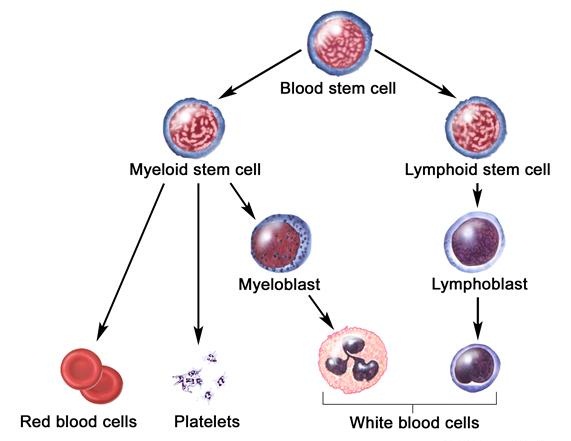
Zones of cell division in animals vary.  In humans (and probably mammals in general), one of the most active areas of cell division is the bone marrow which produces blood cells.  On average, a human turns over about 120 million red blood cells a day.  Having said that the blood stem cells have the potential to become not only red cells but platelets and a variety of white cells.  That is part of the differentiation process, something mentioned earlier.

However, the stem cells need to divide before differentiating.  Mitosis is the solution to this.


Removing Nitrogenous Wastes

Nitrogenous wastes are produced when the amine group of amino acids are removed.  This process is called deamination and take place in the liver.  An example is below.


One of the problems is the toxicity of ammonia.  It needs to be either quickly converted or removed from the body.  Examples of how ammonia is treated by various animals is shown below 


As you can see, most fish don't bother converting the ammonia.  They just excrete it straight into the environment via the gills.  Sharks convert it to urea and use it to regulate their internal fluid levels (not needed for this course).  However, land based and egg laying animals are faced with the challenge of not poisoning themselves while retaining water.  As a result while benefits are gained, it comes at a cost in energy.


Mammals and amphibians use the urea molecule as their solution.  It is about 100 times less toxic than ammonia, and very water soluble.  As a result removal of urea can occur with far less fluid.  In mammals the main organ of excretion is the kidney.

The fluid from the blood is squeezed into part of the nephron called the Bowman's capsule.  From there, it travels along the tubule.  By a combination of active transport, osmosis and the selectively permeable nature of membranes, things that the body needs are returned to the blood and waste salts and urea are removed via the urine stream.


Birds and reptiles also have kidneys.  However the material they excrete is uric acid.  Uric acid has a 2 key characteristics.

  1. It has very low toxicity
  2. It is very insoluble. 

Uric acid causes a great deal of pain to gout sufferers because of its insolubility.  But it means that if you are developing in an egg for a few weeks, the material will solidify before it can poison you.  In addition, it means that very little water is needed to get rid of it as it can be fed into the fecal pellets and voided with that waste.  Pictured below are the wastes of lizard and bird. The uric acid is the white stuff.


Insects on land generally lay eggs and so use uric acid as a means disposing of nitrogen containing wastes.  It is collected by a series of tubules in their bodies called Malpighian tubes.  The uric acid is concentrated and is then fed into the digestive tract where it can be excreted as part of the feces. 


Gas exchange 2 - Countercurrent flow and Plants

Fish gas exchange

Because the amount of oxygen in water is about 1/20th compared to air, fish need a highly efficient means of oxygen extraction. To this end they combine high SA to vol with something called counter-current gas exchange.  


Counter current flow means that the blood and the oxygen flow in opposite directions to each other (shown on the left hand side of the figure below).  If the blood and water flowed in the same direction, it would be called concurrent flow. 

The real benefit of counter current flow is the ability of the blood to always have a slightly lower concentration than the water flowing over it.  This means a concentration gradient is established for longer and more oxygen is removed from the water to the blood for the fish.  If blood and water flowed in the same direction, then the concentrations would be equal at some stage and no further gas exchange could take place. 


Plant Gas exchange

In the leaf, gas exchange structures are the stomates.  These not only allow O2 out and CO2 in,  but also allow minerals to be transported up the plant as transpiration acts as the driving force for the movement of water up the plant.

Stomates in plants that live in warmer drier climates like Australia tend to open in the morning, close down in the middle of the day, and then reopen in the afternoon.  This way, water loss is reduced in the warmest part of the day, but light intensity is not compromised. Stomates are generally on the underside of the leaves away from direct sunlight which also helps with water conservation.


Lenticels are corky areas of loosely packed cells that are located on branches, stems, and the trunks of plants.  They are able to let oxygen into these parts of the plants that cannot perform photosynthesis to be used in respiration and allow CO2 to escape.  


When it comes to the roots of plants, most absorb oxygen through from air spaces within the soil. This is why over-watered plants die, because the roots literally drown.  However, some plants live in areas where air can never get into the soil to allow them to efficiently exchange oxygen and CO2.  The best example of this are mangroves.  To compensate for this they have snorkel like extensions on their roots called pnematophores.

Thursday 28 March 2013

Gas exchange 1 - Mammals, Insects and Amphibians

The essential features of a gas exchange system is as follows:


  1. High surface area
  2. A moist surface to allow gases to dissolve 
  3. Thin structures to allow rapid diffusion into the tissues of the organism
  4. Well supplied with blood vessels

In the case of insects, oxygen is obtained and CO2 removed  via openings in the abdomen.  These are called spiracles   When the air passes into the insects it enters the tracheal tubes and passes down a series of branched tubes called tracheoles.  These small structures have high SA and so allow for gases to be exchanged with the tissue fluid.  The tracheoles are attached to the muscles that pulse, drawing air in and pushing it out.


In the case of amphibians, the lungs are not well developed and do not have sufficient surface area to allow for sufficient gas exchange.  To supplement this amphibians also use their skin as a gas exchange surface.  Recall earlier the need for the surface o be kept moist.  This is the primary reason that a frogs skins must be moist at all times.  

Frogs breath by positive pressure means.  That is they push the air into their lungs by sealing off the nostrils and pushing with their bucal cavity.  So when you see the frogs "chin" moving, it is in the act of breathing.


In humans, negative pressure breathing occurs.  The lungs are elastic and so when the diaphragm pulls them downwards, the pressure inside the lungs is less than the outside air.  As a result, air rushes in.  To exhale the reverse occurs.  Humans have massive surface area inside their lungs.  One problem they would have if their lung surfaces were directly exposed to the environment is rapid dehydration.  To stop this from happening, the lungs are internalized in the chest cavity, and air can only enter and exit via the nose or mouth.  Most of the moisture is trapped before it leaves the body.  However, water losses are inevitable.  This can be seen when you breath on glass or on a cold winters day.


Air travels down the trachea, bronchi and bronchioles until it reaches the alveoli.  This is the site of gas exchange.  The alveoli are shaped like clusters of grapes.  This increases surface area for gas exchange.  As shown in the diagram below, these are also well supplied with blood vessels. 


When people develop emphysema, the alveoli begin to breakdown and reduce the surface area for gas absorption.  This results in the shortness of breath that sufferers have.