Saturday 23 February 2013

The Chemistry of Cells and Organisms


One of the things that you need to be aware of is the way in which organisms obtain their nutrition from.  These terms may be handy.

  1. Photoautotraph - makes own food by using light energy.  Photosynthesis is used by plants and some types of bacteria.
  2. Chemoautrotroph - make own food using the energy of simple chemicals. Bacteria found in deep ocean vents and hot springs are capable of this.
  3. Heterotroph - obtains food by ingesting complex materials from autotrophs or other heterotrophs.


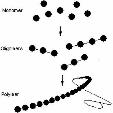
When it comes to nutrients, they can be arranged into a number of categories.  Monomers are chemical groups that are made up of a single functional unit.  These can be assembled into small chains of 2 or 3 units, called oligomers or long chains called polymers. 


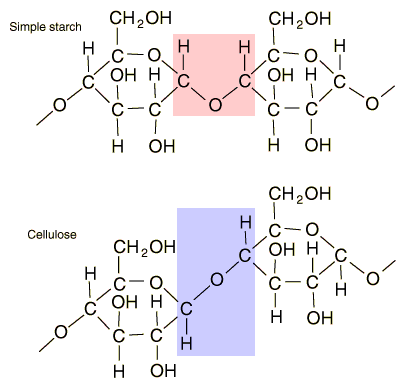
The first class of compounds are carbohydrates (CH2O - carbon + water).  These provide energy when a monomer.  The best known example is glucose.  However, glucose monomers can be assembled into polymers for other uses:

Starch (plants) - longer term energy storage
Glycogen (animals) - longer term energy storage
Cellulose (plants) - material for cell walls, to provide rigidity and strength to plant cells.


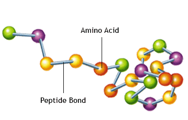
The second class of materials are proteins and amino acids.  Amino acids are the build block for proteins.  Proteins have many functions in a cell, but at this stage we will say that they are used for cell growth and repair.


Other classes of materials include fats and oils.  These are used not only as in cell membranes but also as a long term energy storage because the amount of energy that they have is very high compared to other biological molecules.


Vitamins are a group of small molecules that are made by living things that assist many of the chemical reactions that take place in out cells.

Minerals include salts and more specifically dissolved ions.  They can also assist with chemical reactions, but help regulate the water balance in cells and are involved with activities such as nerve signal transmission and muscle contraction.


Wednesday 20 February 2013

Diffusion and Osmosis

Diffusion is the movement of dissolved solids, liquids and gases from areas of high concentration to areas of low concentration until all the molecules concerned are evenly distributed.  

When this occurs the system is said to be in equilibrium and while molecules still move, they remain evenly spread in the solution.  A good example is using Condy's crystals (potassium permaganate) in water.


When the crystals settle on the bottom of the water and begin to dissolve, you can see that the bottom is concentrated with the coloured permaganate while the dilute solution is at the top.  

This difference in concentration is called the concentration gradient and is required for diffusion to take place.

As time progresses, the random motion of the dissolved ions sees the colour begin to spread (30 min later).  However, it can still be seen that the solution at the base of the beaker is still more concentrated than that at the top.  Given more time, the remaining crystals dissolve, and the permaganate ions eventually move around in the fluid until they are evenly distributed and are at equilibrium. The permaganate ions have diffused from the bottom of the beaker until evenly spread.


In cells, differences are likely to occur between the inside of the cell and its external environment.  If the membrane is permeable to the material it will cross the membrane and establish an equilibrium.


It should be pointed out that the concentration gradient does not always have to from the outside of a cell going in, it can operate in reverse, being more concentrated on the inside of the cell moving out.  2 way flow of substances in opposite directions can occur as shown above.


The movement of water which is driven by concentration gradients is called osmosis.  The reason that water is dealt with separately is because its movement often happens in the opposite direction of other molecules.  

Lets look at our Condy's crystal eaxmple again, but consider BOTH the permaganate ions and the water molecules in the beaker this time.


As the crystals dissolve, the permaganate ion concentration is high at the bottom of the beaker but low at the top.  However, when we think about the water molecules, the concentration of these are high at the top and low at the bottom.  So as the permaganate ions move upwards, the water molecules will be moving downwards to ensure everything is evenly distributed and an equilibrium is reached. 

So while achieving the same result water needs to be considered separately because while it follows the same rules, it seems to act in a counter intuitive way.


In many cells substances may be unable to cross the membrane, and so to try and establish an equilibrium, water must move to do this.  In the figure below, because the sucrose is unable to pass through the membrane, it is up to the water to move by osmosis in an effort to try and dilute it and establish an equilibrium.


Cells can behave in a number of ways to this changing amounts of water relative to the concentrations of water inside the cell membrane.


In (a), the cells have been placed in a hypertonic solution.  This means that the amount of water inside the cell is greater than the outside environment.  This happens when a high concentration of solutes are in the water surrounding the cells.  As a result water leaves the cell causing shriveling or plasmolysis.

In (), the cells have been placed in a isotonic solution.  This means that the amount of water inside the cell is equal to the outside environment.   As a result water leaves  and enters the cell at the same rate leaving it unchanged.

In (c), the cells have been placed in a hypotonic solution.  This means that the amount of water inside the cell is less than than the outside environment.  This happens when a very low concentrations of solutes are in the water surrounding the cells.  As a result water enters the cell causing animal cells to burst, or plant cells to become turgid.  The plant cell wall pushes back on the membrane, making it rigid, but stopping it from breaking.  This is how lettuce and celery can be revived and made crisp again after storage.


Thursday 14 February 2013

Cell Membranes


The cell or plasma membrane surrounds the cell and protects the cell contents from the external environment.  The main component of the membrane is the phospholipid.  The phosphate group (orange circles) sticks itself into the watery world of the environment or cytoplasm, while the water hating oily lipids  (black lines attached to orange circles) organise themselves so that they face each other away from the water. In doing so they form a 2 molecule thick bilayer.  This acts as a pretty effective barrier for the cells to keep their contents in and the environment out.


However, there are things that the cell needs to get in (like nutrients) and out (like wastes).  To help this the membrane incorporates proteins into this bilayer. These let materials cross the cell membrane according to their shape. If the materials are the right shape, they can pass through the channel and enter or exit the cell. If not, they stay where they are.

Proteins also act as way of cells recognising each other (important for the immune response not killing your own cells).

The other structures on the cells include carbohydrates and cholesterol.  Carbohydrates also help in cell recognition (blood typing being a good example) while cholesterol helps keep the membrane fluid.

When we draw the membrane in a 3 D manner, things get complex.


Basically the structure of the membrane is made up of many small pieces arranged together to create a functional unit.  This is like a mosaic picture, where the picture is made up of many fragments.  Because membranes have to be fluid to work, the model of the membrane is called the fluid mosaic model.


Because the membrane now only lets certain things though now, it is called a specifically permeable or semi-permeable membrane.

Shown below are examples of an impermeable (nothing gets through, on theleft) and fully permeable membrane (everything gets through on the right).


In the example below here, note that only 1 of the 2 substances gets through.  The membrane selected the red circles to pass through, but not the purple ones.  This is an example of selectively permeable. 

Wednesday 13 February 2013

Cell organelles and their function

Looking at the dot point you can see that the list of organelles that yoou are expected to see under a light microscope and electron micrograph is fairly comprehensive.  It includes:

mitochondria, chloroplasts, vacuoles, Golgi bodies, cell wall, lysosomes, endoplasmic reticulum, ribosomes, nucleus, nucleolus and cell membranes.


Under a light microscope you can expect to see the in an animal cell; the cell membrane, the nucleus and the cytoplasm.


Under a light microscope you can expect to see the following for a plant cell; the cell membrane, the nucleus, cell wall, chloroplasts, vacuole and the cytoplasm. I should point out that you are unlikely to see them all in a single specimen, but you should be able to see some of these on any given slide.


In an electron microscope image the following are generally visible in animal cells; mitochondria, Golgi bodies,  lysosomes, endoplasmic reticulum, ribosomes, nucleus, nucleolus and cell membranes.  Not all are visible in this image however.


An electron micrograph of a plant cell will contain the same as an animal cell but will also have in addition the chloroplasts, vacuole and cell wall.  Interestingly animal cells have vacuoles but they are very small and not visible on any image you will see.  Therefore we consider vacuoles absent from animal cells


As for the functions of these cell organelles, I am leaving it up to you to figure out what they are.


Technology and its impact on our understanding of cells

There have been a number of technological developments that have advanced out understanding of cells:

1.  The invention of the microscope.  Without this, nothing would have started.

2.  The use of dyes and stains.  These were a byproduct of the fabric industry in the 2nd half of the 19th century.  When people tried them out on specimens mounted on the microscopes, they found that they could see structures that were previously invisible.


3.  The use of oil immersion and light manipulation (eg. phase contrast).  Oil immersion pushed the magnification limits of the microscope out to 1000 X while phase contrast techniques allowed for better contrast of specimens highlighting structures either unseen or unnoticed.


4.  Electron Microscopes.  Because they used a beam of electrons at a much shorter wavelength than visible light, magnification and resolution was greatly enhanced.  


When comparing the advantages of light and electron microscopes, you tend to find that the disadvantage of one is the advantage of the other.

Advantages of light microscopes include; price, ease of use, live specimens can be mounted, images can be seen directly in colour.

Advantages of electron microscopes are; extremely high resolution and magnification (which trumps a lot of the disadvantages)

Disadvantages of light microscopes may be; limits to magnification and resolution (a biggie), some training to use stains, lighting and oil immersion techniques needed.

Disadvantages of electron microscopes are; costly, specialised training required to use, specimens are cannot be live, images will be in black and white unless computer enhanced. 


Monday 11 February 2013

The Cell Theory


In 1838 Schlieden and Schwann first proposed the cell theory.  They stated that:

1.  All living things are made of cells
2.  The cell is the basic unit of all living organisms.

In 1855 Virchow added another rule.  It was:

3.  All cells come from preexisting cells.


Who were the other players in this?

Robert Hooke.  He first coined the term cell in 1665 while sketching a slice of cork under his newly built compound microscope.


Redi - His observations linking flies with maggots in rotting meat led to the beginning of the end for spontaneous generation.  His work was later verified by Pasteur who made similar observations with yeast turning grape juice into wine, and then bacteria spoiling the wine.


Leeuwenhoek - observed living single-celled in pond water under his microscopes.  He also observed that blood of a variety of organisms had cells in them.

Robert Brown - his observations showed that there was potential complexity within the cells by observing and naming the cell nucleus.  While it may have been observed earlier, Brown was one of the first to publish these into scientific literature. 


What evidence supports cell theory?

Observation with the microscope.  As time went on and better light microscopes were developed, the key characteristics of all living things certainly followed the first 2 rules.  All living things had cells in them and the smallest living thing is made up of cells.

With the discovery of dyes that stained the nucleus and other cell parts, rule 3 was verified by Walther Fleming in 1879.